Locations:
Advertisement
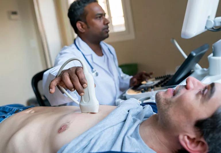
An ultrasound of your heart allows doctors to see it pumping blood
If your primary care doctor or cardiologist has ordered a transthoracic echocardiogram, don’t fret – it’s not as intimidating as it might sound.
Advertisement
Cleveland Clinic is a non-profit academic medical center. Advertising on our site helps support our mission. We do not endorse non-Cleveland Clinic products or services. Policy
Called an “echo test” for short, it’s a painless test that’s noninvasive and doesn’t use ionizing radiation. What it does do is equip your doctors with critical information about how your heart is functioning that can help them diagnose and treat disease, or confirm that things are working just fine.
Here’s what’s in store.
An echo test is an ultrasound of the heart. It uses high-frequency sound waves to produce images of the heart’s valves and chambers so that doctors can see how your heart is functioning.
“We look at all the chambers of the heart, the heart size, the heart function, how strong the heart is beating, and pressures within the heart and across the valves,” says cardiologist Rhonda Miyasaka, MD. “We can also see how well the valves are functioning – for example, we can see if they’re leaking or if they’re having trouble opening.”
The test has been around for a long time, but the quality of the images that doctors can get with it continues to improve. “We can now see three-dimensional pictures of the heart and valves which can be very helpful, and we have newer, more detailed technologies to measure heart function,” Dr. Miyasaka says.
Echo tests are often combined with Doppler imaging technology to show how blood is flowing across the heart’s valves.
Doctors might want to see an echocardiogram to investigate signs or symptoms of heart diseases, like shortness of breath, chest discomfort or swelling in the legs.
They might also order an echocardiogram if something abnormal, like a heart murmur, is detected during an exam. It can help them see if there’s been damage to the heart muscle, or a problem with a valve, Dr. Miyasaka says.
Depending on why the echo test is being ordered, your doctor might also order other diagnostic tests like blood work or an electrocardiogram.
There’s no special preparation required – you can take any medication and eat and drink as usual before the test. You will typically go to an outpatient facility, and it should take no more than an hour.
During the echo test, you’ll be asked to put on a hospital gown. You’ll lie on an exam table, and a sonographer or ultrasound tech will put some gel on the end of an ultrasound wand and move it along your chest. The gel might be a little cold, but otherwise you should not feel any major discomfort during the test.
In some cases, doctors might need to inject a contrast agent into your bloodstream in order to see the borders of the heart better.
Advertisement
If your test was ordered by a cardiologist who can read his or her own echo test results, the doctor should be able to figure out pretty quickly if there’s anything abnormal going on with your heart. If it’s been ordered by a non-cardiologist, they should get the official results report within a day.
Possibly. “If someone has a known cardiac condition, then echo is one of our gold standard tools for following that heart condition,” Dr. Miyasaka says. So, someone who is diagnosed with a chronic heart condition will have multiple echos over their lifetime to help doctors monitor the progression of the disease and determine the best course of treatment.
An additional echocardiogram also might be ordered if you switch health systems or are referred to a new doctor. “Echo is very dependent on the skill of the sonographer, who is specially trained to do cardiac ultrasound,” Dr. Miyasaka says. If your new doctor feels like the sonographers on his or her team can take more detailed, high-quality images, they may order a new test.
If they need a closer look at how your valves are functioning, doctors may order a transesophageal echocardiogram. Patients are sedated during this procedure, which uses a special ultrasound wand that is inserted down the throat and into the esophagus, right behind the heart.
There’s also something called an exercise stress echocardiogram that’s used to detect problems with the arteries that supply blood to your heart muscle. It’s the same as a traditional echocardiogram, except the sonographer will take images of the heart before and after a brief period of exercise on a treadmill or stationary bike.
Advertisement
Learn more about our editorial process.
Advertisement

Factors like temperature, energy levels and sleep quality play a role in determining whether working out in the morning or evening is best for you

Obesity, age and preexisting heart conditions can all raise your risk of cardiovascular disease during pregnancy

Xylitol in processed food can increase risk of heart attack and stroke — but there’s no danger in xylitol in oral care products

If your provider has ruled out a serious cause, you can treat chest pain at home with antacids, inhalers or anti-inflammatory medications

Walking is a great goal, but how many steps are best for you depends on factors like your fitness level and age

Research shows a strong association between rheumatoid arthritis and heart issues

Eating more natural, whole foods can lower your risk of heart and cardiovascular diseases

First things first — slowly sit or lie down

Focus on your body’s metabolic set point by eating healthy foods, making exercise a part of your routine and reducing stress

PFAS chemicals may make life easier — but they aren’t always so easy on the human body

While there’s little risk in trying this hair care treatment, there isn’t much science to back up the claims