Locations:

One medical test, two different parts
If you’re pregnant or need a medical diagnosis, your provider might order an ultrasound. Or is it a sonogram? Many people use these terms interchangeably, but they’re not necessarily the same thing. And technically, you can think of them as two sides of the same coin.
Advertisement
Cleveland Clinic is a non-profit academic medical center. Advertising on our site helps support our mission. We do not endorse non-Cleveland Clinic products or services. Policy
Radiologist Albert Parlade, MD, clears up the confusion around sonograms versus ultrasounds, and how and why they’re used for various circumstances.
Medical terms can be confusing. You’ve probably heard people say they’re “getting a sonogram” or “need an ultrasound done.” But what do these terms actually mean, and is one better than the other?
“Sonogram and ultrasound are two different parts of the same medical test,” says Dr. Parlade. “The word ‘ultrasound’ refers to sound waves used during an ultrasound exam, which is a medical imaging test. These waves are higher than what humans can hear, but they can create images of body tissues.”
A sonogram, then, is the picture created by the ultrasound machine.
“Think of the ultrasound machine as your camera, and the sonogram as the photograph you get from the camera,” clarifies Dr. Parlade.
And in the case of a prenatal ultrasound, you might receive a printout picture of the sonogram after your exam.
An ultrasound exam collects images of soft tissues inside your body. Most people associate ultrasound with pregnancy, but it’s also used for many other medical reasons.
“Ultrasound exams help providers diagnose or rule out many conditions,” says Dr. Parlade. “They’re noninvasive, painless and don’t use radiation.”
Advertisement
An ultrasound exam can spot growths or abnormalities inside of your body. It can also clearly show and measure fluids.
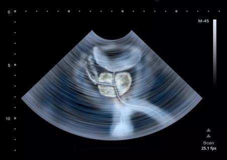
“Ultrasound waves go straight through water, so water appears black on a sonogram,” explains Dr. Parlade. “If the fluid is infected or contains blood, it shows up in shades of gray. An ultrasound also highlights textures and details on your tissues and organs.”
Providers use ultrasound exams to view:
But an ultrasound can’t see everything. It’s usually not the best test to view bones.
“Bones block sound waves, so they don’t show up well on an ultrasound exam,” explains Dr. Parlade. “We usually perform an X-ray if we need to get a picture of a bone. Ultrasound also cannot see structures inside your joint, such as cartilage.”
In addition, an ultrasound isn’t always the first exam choice for organs that contain air, such as your small or large intestine (although an endoscopic ultrasound can be used to examine the inside of your digestive system).
“Air appears bright white on a sonogram and obscures the image,” he adds. “We typically use other tests, like a CT scan or MRI, if we need an image of the bowel.”
An ultrasound isn’t just a standalone medical test. Providers also use this technology to guide minimally invasive procedures, like a breast biopsy.
An ultrasound machine can generate real-time sonograms during a biopsy or cyst drainage. The sonogram allows your provider to see the exact location of the needle or tool without a large incision. Providers also use ultrasound guidance during IVF (in vitro fertilization) to guide an embryo into the uterus.
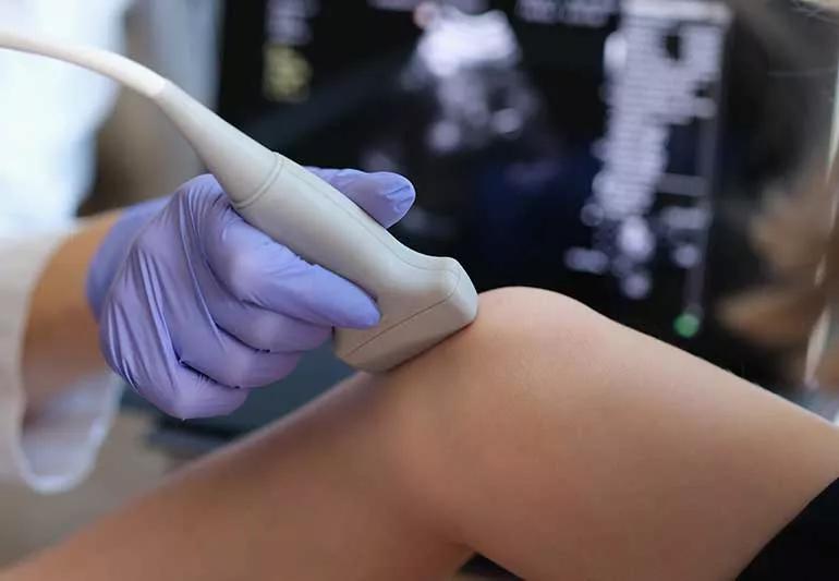
If you need an ultrasound exam, you see a sonographer. This medical professional specializes in using an ultrasound machine to collect clear images.
The sonographer places an ultrasound probe on your skin or inside a natural opening of your body, like your vagina or anus. During the test:
“Your sonogram is a real-time, moving image that appears on a computer screen during your ultrasound exam,” says Dr. Parlade. “During your test, the sonographer periodically freezes live images to create snapshots of important information. They may also save video loops to show movements of a fetus or fluid.”
Advertisement
It’s common to get a printout of your sonogram after a pregnancy ultrasound exam. This is the black-and-white image that parents often save. Your obstetrician views your sonogram to check the health and well-being of the developing fetus.
If you need a sonogram for another medical reason, you may not receive a printout afterward unless you ask for one. But a radiologist will view and interpret your sonogram images and video loops. Usually, the radiologist sends a report to your referring provider with a diagnosis or other information based on your results.
Whether you call it an ultrasound or a sonogram, you’re getting the same imaging test done. Fortunately, you don’t have to worry about whether one is better than the other.
If you have questions about an ultrasound or any medical procedure, talk to your healthcare provider. They can tell you why you need the test and what they’re looking for. When you’re empowered with that information, you can make better, informed decisions for your own medical care.
Advertisement
Learn more about our editorial process.
Advertisement

CTs and MRIs use different technologies to show different things — neither is necessarily ‘better’ than the other

From faster diagnoses to less paperwork, healthcare providers are using artificial intelligence to help take better care of you

While an ultrasound shows your muscles and tendons, an MRI also shows your joint cartilage, bones and heart chambers
This noninvasive procedure can help aging skin by boosting collagen production
High-intensity focused ultrasound (HIFU) provides new treatment options

A closer look at getting a SIS

People with PKU need to avoid high-protein foods, like meat, dairy, legumes and whole grains

Introverts refuel with quiet time, while extroverts thrive on connection

Wearing a scarf, adjusting your outdoor activities and following your asthma treatment plan can help limit breathing problems

Your diet in the weeks, days and hours ahead of your race can power you to the finish line

When someone guilt trips you, they’re using emotionally manipulative behavior to try to get you to act a certain way